|
RESEARCH |
|
|
|
CytoJournal 2008,
5:4 |
Fine needle aspiration biopsy of intraoral and oropharyngeal mass lesions
Husain A Saleh1, Lewis Clayman2, Haitham Masri3
1 Department of Pathology, Sinai-Grace Hospital/Detroit Medical Center, Wayne State University, Detroit, Michigan, USA 2 Department of Pathology, Sinai-Grace Hospital/Detroit Medical Center, Wayne State University, Detroit; Department of Oral-Maxillofacial Surgery, W. Beaumont Hospital, Royal Oak; Department of Head & Neck Surgery, University of Michigan, Ann Arbor, Michigan, USA 3 Department of Otolaryngology/Head and Neck Surgery, Oakwood Hospital and Medical Center, Dearborn, Michigan, USA
Date of Submission | 15-Dec-2007 |
Date of Acceptance | 28-Mar-2008 |
Date of Web Publication | 28-Mar-2008 |
Correspondence Address: Husain A Saleh Department of Pathology, Sinai-Grace Hospital/Detroit Medical Center, Wayne State University, Detroit, Michigan USA
 Source of Support: None, Conflict of Interest: None  | Check |
DOI: 10.1186/1742-6413-5-4
Background: Fine needle aspiration (FNA) biopsy has been rarely used in oral and oropharyngeal lesions. The goal of this study was to assess the value and accuracy of FNA biopsy in the diagnosis of oral and oropharyngeal lesions particularly in regards to discriminating benign from malignant tumors. Methods: Sixteen cases of FNA biopsies obtained of various intraoral and oropharyngeal masses or lesions performed at our institution during the eight-year period from 1998 to 2006 were retrospectively reviewed. The aspiration cytologic diagnoses were correlated with the histologic examination of the corresponding resected lesions. Results: Sixteen cases of intraoral lesions evaluated by FNA biopsies during the period of 1998-2006 were reviewed. The sites of involvement were: lip [1], maxillary sinus [3], pharynx/oropharynx [5], floor of mouth [4], buccal mucosa [2] and peritonsillar area [1]. Patients' age ranged from 30 to 87 with an average of 54 years. Male to female ratio was 1:3. Cytologically, 7 cases were diagnosed as suspicious/malignant, and 9 cases as benign (including 6 benign neoplasm, 1 atypical, and 2 reactive or "descriptive"). Fifteen cases had corresponding surgical resection for histologic examination, of these, 9 cases were interpreted as malignant, and 6 as benign. There were no false positive diagnoses of malignancy on FNA. Two cases were interpreted as benign or atypical cytologically, but were found to be malignant on histologic examination. Conclusion: FNA biopsy of intraoral and oropharyngeal masses is a valuable procedure for the initial evaluation of various lesions. It provides helpful information about these lesions and avoids hasty or unnecessary surgical biopsy. It is a rapid and relatively noninvasive procedure. Furthermore, aspiration biopsy is an important tool in the diagnosis and management of these lesions, both neoplastic and non-neoplastic, and can be sometimes complemented by ancillary studies for more accurate interpretation. However, its sensitivity in the diagnosis of malignancy is lower than that of histologic samples. This is probably due to the superficial nature and small size of these lesions, the limited space for maneuvering the needle and difficulty in immobilizing the lesion to obtain adequate samples, rather than to interpretation or inherent limitations of the technique itself.
How to cite this article: Saleh HA, Clayman L, Masri H. Fine needle aspiration biopsy of intraoral and oropharyngeal mass lesions. CytoJournal 2008;5:4 |
Introduction | |  |
A wide variety of benign and malignant tumors and non-neoplastic lesions can arise in the intraoral and oropharyngeal areas [1,2]. Squamous cell carcinoma is the most common malignancy, and pleomorphic adenoma of minor salivary glands is the most frequently encountered benign tumor, which usually involves the palate [3-9]. Clinically, cancer of this area may present with pain, bleeding or mass lesion. Traditionally, these masses have been evaluated by surgical biopsy or exfoliative cytology. FNA biopsy has proved to be safe, economic, simple and accurate in the evaluation of various organs including the head and neck [10-12]. However, aspiration biopsy studies of the intraoral and oropharyngeal masses are few and limited [13-16].
In this study, we retrospectively reviewed 16 cases of intraoral and oropharyngeal lesions sampled by FNA biopsy with particular attention to the cytologic features of specific tumors, diagnostic accuracy and cytologic- histologic correlation.
Materials and methods | |  |
Sixteen cases of intraoral and pharyngeal masses and lesions that were sampled by FNA biopsies during the period of 1998-2006 were retrieved from the archives of the pathology department of the Detroit Medical Center, Detroit, Michigan. There were 4 men and 12 women with a male: female ratio of 1:3. They had wide age range from 30 to 87 years with an average age of 54 year old. The pharynx- oropharynx was the most common site of involvement (5 cases). Other sites included the maxillary sinus [3], floor of mouth [4], buccal mucosa [2] and lip and paratonsillar (1 for each). The FNA biopsies were performed mostly by the head & neck surgeons with on-site evaluation for adequacy and triage by cytopathologist, cytopathology fellow or experienced cytotechnologist on most cases. The aspirates were performed, consistently and similarly by all the physicians, according to the standard procedure using 22-23 gauge needles connected to a 10 c.c. plastic syringe and sometimes mounted on Cameco aspiration handle (Precision Dynamics Corp., Burbank, California, USA). After localizing and immobilizing the mass, the aspiration needle was passed through the lesion 2 or 3 times in each case. Air- dried Diff-Quik stained smears were made for immediate evaluation, the remaining smears were alcohol- fixed and stained with Papanicolaou method. The syringe and needle were also rinsed in cytofixative and cell blocks were made in 14 cases whenever sufficient material permitted.
Of the 16 FNA cases, 15 had subsequent corresponding surgical specimens, either a biopsy or surgical resection.
Results | |  |
[Table 1] shows the demographic, cytologic and histologic findings of all patients. The average patient's age was 54 years (range 30-87). There was an obvious female predilection with a male: female ratio of 1:3 (4 men and 12 women). The distribution of the lesions varied, but pharynx/oropharynx was the most common location [5]. Other sites included: upper lip mucosa [1], left peritonsillar area [1], maxillary sinus [3], floor of mouth [4], and buccal lesions [2]. The most common clinical presentations were pain, bleeding and a mass lesion.
Cytologically, nine cases were diagnosed as benign and included: epithelial/myoepithelial lesion [1], granular cell tumor [2], pleomorphic adenoma [2], squamous cyst [1], reactive/descriptive [2], and atypical squamous cells [1]. Of the two reactive/descriptive cases, one was called reactive lymphoid population and was of a swelling of peritonsillar area confirmed histologically to be reactive lymphoid hyperplasia; and the second was called "benign fibrous and epithelial cells" and was of a right maxillary sinus mass confirmed to be unicystic ameloblastoma. The case of right lateral oropharyngeal lesion called "atypical squamous cells" on FNA turned to be invasive squamous carcinoma on surgical resection. On the other hand, seven cases were interpreted as suspicious or malignant and included: adenocarcinoma [1], sarcoma [1], carcinoma NOS [1], mucoepidermoid carcinoma [1], plasmacytoma (!) and squamous cell carcinoma [2].
Histologically, 15 of the 16 FNA cases had corresponding histologic follow up. [Table 1]. Of these 6 of 9 cytologically diagnosed benign lesions were also confirmed to be benign (one had no histologic follow up). Two case, however, were found to be malignant (2 false negative FNA cases). one was a left peripharyngeal nodule cytologically called epithelial-myoepithelial lesion and was found to be metastatic breast carcinoma on resection. The second case, lateral oropharynx, was called atypical squamous cells and was found to be squamous cell carcinoma on surgical resection.
Importantly, all 7 cases called malignant on aspiration cytology were also found to be malignant on surgical specimens, with no false positive diagnoses. All of the histologically confirmed benign cases were also diagnosed as benign on FNA cytology (specificity 6/6; 100%). However, as mentioned earlier, we failed to identify two malignant cases on FNA cytology, one was interpreted as benign and one as atypical, which results in sensitivity of (7/9; 77.7%) for malignant diagnosis (2 false negative FNA cases) [Table 2]. On further review, these two cases were found to have on-site evaluation done by cytopathology fellow in training.
Lymphoid hyperplasia
The FNA smears of the peritonsillar swelling revealed cellular aspirate with singly dispersed mixed population of lymphocytes, plasma cells and scattered histiocytes. Flow cytometry studies confirmed polyclonal lymphoid tissue.
Ameloblastoma
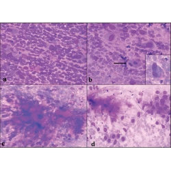
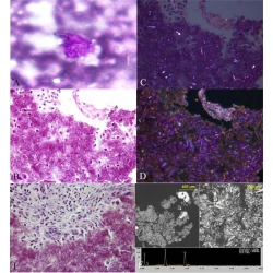
The FNA smears of right maxillary sinus mass revealed palisading groups of epithelial cells admixed with mesenchymal elements. The cells showed low grade atypia, [Figure 1a,b,c].
Sinonasal undifferentiated carcinoma
The FNA smears and cell block showed groups and 3-dimentional clusters of atypical malignant epithelial cells with irregular hyperchromatic nuclei and small nucleoli [Figure 2a,b].
Pleomorphic adenoma
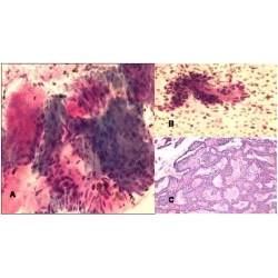
The aspirate smears showed biphasic population of epithelial cells and mesenchymal fibrillary myxoid substance. The round to oval epithelial cells were free or mixed within the mesenchymal tissue [Figure 3a,b,c].
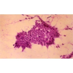
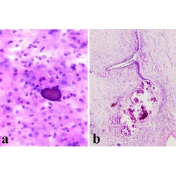
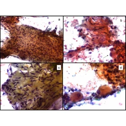
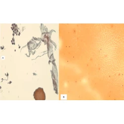
The right peripharyngeal FNA case incorrectly interpreted as granular cell tumor showed loose clusters or singly dispersed round to polygonal cells with abundant dark eosinophilic cytoplasm and occasional peripheral nuclei [Figure 4A]. Interestingly, the resection specimen of this mass revealed adult rhabdomyoma [Figure 4B] that was confirmed by immunostains of myoglobin and myogen (interpretation error of benign tumor). The second FNA case of right mandibular mass showed loose groups of polygonal cells with well demarcated eosinophilic granular cytoplasm and central round nuclei with small nucleoli. The cells were strongly positive for S-100 protein immunostain performed on the cell block confirming the diagnosis of granular cell tumor [Figure 5a,b].
Mucoepidermoid carcinoma
The FNA smears of the left mandibular area mass displayed mixture of mucous cells (including some goblet cells), squamous cells and few polygonal cells with mucous material in the background. All cells had mild atypia. The cell block section was helpful in revealing fragments with intimate mixture of atypical squamous and glandular cells including goblet cells, consistent with low grade mucoepidermoid carcinoma [Figure 6a,b].
Squamous cell carcinomas
The aspirate smears showed three dimensional groups and discohesive clusters of atypical squamous cells with varying degrees of keratinization and nuclear atypia. Pleomorphic, hyperchromatic and elongated nuclei were seen, [Figure 7a,b].
The FNA smears of the left posterior buccal mass showed cellular sample with many somewhat atypical singly dispersed plasmacytes. Size variation and occasional binucleated plasma cells were present. This was diagnosed as plasmacytoma and confirmed by immunostains for CD 138, [Figure 8a,b].
The right buccal lesion aspirate displayed many anucleated squames, some spindly or bland benign squamous cells and keratin material consistent with benign squamous cyst. The resected specimen also showed retention squamous cyst with benign mature squamous epithelial lining.
Discussion | |  |
The oral and pharyngeal areas can be home for a wide variety of benign and malignant tumors and non-neoplastic lesions. Squamous carcinoma is the most common malignancy [1], but adenoid cystic carcinoma and mucoepidermoid carcinoma of minor salivary glands are also common in this region. On the other hand, pleomorphic adenoma arising from minor salivary glands is the most frequently encountered benign tumor often found in the palate [3-9]. Usually, these patients present with local pain, bleeding or a mass lesion. Surprisingly, many of the patients with oral and oropharyngeal cancers have advanced disease at the time of diagnosis [1,2]. Surgical biopsy is the traditional method for evaluating these lesions but this method may be inconvenient, painful, and costly due to hospitalization and may leave underlying tissue damage. Also, exfoliative or scrape cytology has been attempted, but this method has high false negative rates up to 37%. It was most useful for superficial epithelial lesions but not subepithelial lesions such as salivary gland tumors and lymphomas [2,13]. Over the years, fine needle aspiration (FNA) biopsy cytology has been found to be very useful, simple, cost effective and accurate in assessing and diagnosing various neoplastic and non-neoplastic lesions of many body sites including especially the head and neck [10]. However, the potential, value and accuracy of this procedure in evaluating intraoral and oropharyngeal masses has been studied only rarely, and only a few reports were found in the English literature on this subject [1, 2, 17-23].
In this study our goal was to investigate the ability of FNA biopsy procedure to accurately diagnose oral and pharyngeal lesions, and to address the cytologic-histologic correlation. We identified 16 cases of this area that underwent FNA biopsy during the period of 1998-2006. Overall, cytologic diagnosis correlated with histologic findings with regards to benign vs. malignant in 13 of 15 cases with accuracy of 86.6% (one case had no tissue). One case of right maxillary sinus was interpreted as "benign, fibrous and epithelial cells" on FNA, and was found to be ameloblastoma on resection (case 3). Another case of right parapharyngeal mass was diagnosed as "suggestive of granular cell tumor" on FNA, but was actually an adult rhabdomyoma on resection (case 7). Importantly, all malignant cytologic diagnoses had a corresponding malignant diagnosis on histology (7 of 7) with no false positive.
Few previous studies addressed the accuracy of FNA biopsy in the diagnosis of oral and oropharyngeal lesions. Scher et al in their study of 93 FNA cases of oral and oropharyngeal lesions, had no false positive diagnoses, but had seven false negative diagnoses [13]. Based on these results and their overall findings, they recommended FNA biopsy for the evaluation of intraoral/parapharyngeal lesions considering its advantages over open biopsy including greater patient comfort, avoidance of general anesthesia, low risk of infection or tissue damage and rapid diagnosis.
In our study, however, 2 cases interpreted cytologically as benign or atypical were actually malignant and later confirmed by tissue examination (false negative diagnoses). The first case of a left oropharyngeal mass revealed what looked like two types of cells: epithelial and myoepithelial suspected to arise from a minor salivary gland. However, the resected specimen showed, surprisingly, metastatic carcinoma of a breast origin. Retrospectively, the cellular pleomorphism (rather than two cell population), immunostains and a history of breast cancer, should have raised the index of suspicion for metastatic breast carcinoma. Finally, the smears of the right lateral oropharynx lesion showed loose discohesive clusters and small irregular tissue fragments of mildly atypical squamous cells. On review, attention to scattered atypical squamous cells with hyperchromatic irregular wrinkled nuclei and considering the presence of a mass lesion may have been good clues to suspect squamous cell carcinoma.
False negative cytologic diagnosis may be due to low cellularity or non-representative sample of the actual lesion. In addition, clinically, some of these lesions are relatively small and superficial making it difficult to reach for precise sampling. They can also be technically difficult to immobilize for making the "back-and -forth" motion in the lesion for ideal aspiration sampling. This emphasizes the significance of correlating the cytologic diagnosis with the clinical history, and if clinical suspicion exists after negative FNA, further investigation should be carried out. Other studies had similar findings and suggested that false negative FNA cytology is most likely related to inadequate specimen or sampling error [15].
One of the malignant cases in our series was a sinonasal undifferentiated carcinoma of the left maxillary sinus and ethmoid recess, which was recognized on FNA as carcinoma, NOS. This rare tumor can be mistaken cytologically with other malignancies and may require immunostains for correct subclassification [12].
FNA biopsy of oral and pharyngeal lesions was shown to be sensitive (93%) and specific (86%) in the study by Shah et. al. Moreover, this technique had advantages over open biopsy with reduced risk of complications such as bleeding, infection and scarring, and ability to identify variety of reactive, benign and malignant lesions [14]. FNA also enables us to triage the specimen during sampling by immediate microscopic evaluation. Cases suspected to be lymphoma or infections can be submitted for flow cytometry evaluation or culture studies, respectively. This triage could help in lowering expenses by avoiding the need for other procedures such as additional samples for culture studies or biopsy. Performing FNA biopsy as initial diagnostic evaluation, rather tissue biopsy, can also save money by reducing in-hospital patient's expenses through eliminating operating room surgical intervention to obtain tissue samples of oral and oropharyngeal lesions.
Conclusion | |  |
In summary, we have demonstrated from our experience of this relatively small series, that FNA biopsy of intraoral and oropharyngeal lesions is a valid procedure and is indeed an important method for the evaluation of these lesions. This technique is simple, financially acceptable and inexpensive, convenient and comfortable to the patient, and above all, can offer a rapid and accurate diagnosis. False negative cases are usually due to inadequate sampling and FNA should be repeated whenever the clinical findings are suspicious for malignancy. Ancillary studies such as immunostains and flow cytometry can greatly help in further classification of the tumors.
Competing interests | |  |
The author(s) declare that they have no competing interests.
Acknowledgements | |  |
The authors would like to greatly thank Mrs. Heidi Staples for her clerical and technical support in conducting this study.[23]
References | |  |
1. | Cramer H, Lampe H, Downing P: Intraoral and transoral fine needle aspiration. A review of 25 cases. Acta Cytol 1995, 39 (4) : 683-8. |
2. | Das DK, Gulati A, Bhatt NC, Mandal AK, Khan VA, Bhambhani S: Fine needle aspiration cytology of oral and pharyngeal lesions. A study of 45 cases. Acta Cytol 1993, 37 (3) : 333-42. |
3. | Buchner A, Merrell PW, Carpenter WM: Relative frequency of intra-oral minor salivary gland tumors: a study of 380 cases from northern California and comparison to reports from other parts of the world. J Oral Pathol Med 2007, 36 (4) : 207-14. |
4. | Hughes KV 3rd, Olsen KD, McCaffrey TV: Parapharyngeal space neoplasms. Head Neck 1995, 17 (2) : 124-30. |
5. | Yih WY, Kratochvil FJ, Stewart JC: Intraoral minor salivary gland neoplasms: review of 213 cases. J Oral Maxillofac Surg 2005, 63 (6) : 805-10. |
6. | Cerulli G, Renzi G, Perugini M, Becelli R: Differential diagnosis between adenoid cystic carcinoma and pleomorphic adenoma of the minor salivary glands of palate. J Craniofac Surg 2004, 15 (6) : 1056-60. |
7. | Gross M, Maly B, Goldfarb A, Eliashar R: Basal cell adenocarcinoma in a buccal minor salivary gland. Acta Otolaryngol 2004, 124 (2) : 213-6. |
8. | Aydin E, Turkoglu S, Ozen O, Akkuzu B: Mucinous cystadenocarcinoma of a minor salivary gland in the upper lip: case report. Auris Nasus Larynx 2005, 32 (3) : 301-4. |
9. | Waldron CA, el-Mofty SK, Gnepp DR: Tumors of the intraoral minor salivary glands: a demographic and histologic study of 426 cases. Oral Surg Oral Med Oral Pathol 1988, 66 (3) : 323-33. |
10. | Stanley MW: Selected problems in fine needle aspiration of head and neck masses. Mod Pathol 2002, 15 (3) : 342-50. |
11. | Stewart CJ, MacKenzie K, McGarry GW, Mowat A: Fine-needle aspiration cytology of salivary gland: a review of 341 cases. Diagn Cytopathol 2000, 22 (3) : 139-46. |
12. | Helsel JC, Bardales RH, Mukunyadzi P: Fine-needle aspiration biopsy cytology of malignant neoplasms of the sinonasal tract. Cancer 2003, 99 (2) : 105-12. |
13. | Scher RL, Oostingh PE, Levine PA, Cantrell RW, Feldman PS: Role of fine needle aspiration in the diagnosis of lesions of the oral cavity, oropharynx, and nasopharynx. Cancer 1988, 62 (12) : 2602-6. |
14. | Shah SB, Singer MI, Liberman E, Ljung BM: Transmucosal fine-needle aspiration diagnosis of intraoral and intrapharyngeal lesions. Laryngoscope 1999, 109 (8) : 1232-7. |
15. | Castelli M, Gattuso P, Reyes C, Solans EP: Fine needle aspiration biopsy of intraoral and pharyngeal lesions. Acta Cytol 1993, 37 (4) : 448-50. |
16. | Domanski HA, Akerman M: Fine-needle aspiration cytology of tongue swellings: a study of 75 cases. Diagn Cytopathol 1998, 19 (3) : 229-34. |
17. | Frierson HF Jr, Covell JL, Mills SE: Fine needle aspiration cytology of atypical carcinoid of the lung. Acta Cytol 1987, 31 (4) : 471-5. |
18. | Pisharodi LR: Low grade papillary adenocarcinoma of minor salivary gland origin. Diagnosis by fine needle aspiration cytology. Acta Cytol 1997, 41 (5) : 1407-11. |
19. | Baehner F, Sudilovsky D: Fine needle aspiration cytology of intraoral epithelioid hemangioma. A report of two cases. Acta Cytol 2003, 47 (2) : 275-80. |
20. | Watanabe K, Ono N, Saito K, Saito A, Suzuki T: Fine-needle aspiration cytology of polymorphous low-grade adenocarcinoma of the tongue. Diagn Cytopathol 1999, 20 (3) : 167-9. |
21. | Kawahara A, Harada H, Yokoyama T, Kage M: Cytomorphologic features of a polymorphous low grade adenocarcinoma of the palate. A report of 2 cases with immunocytochemistry. Acta Cytol 2003, 47 (6) : 1127-30. |
22. | Sahai K, Kapila K, Dahiya S, Verma K: Fine needle aspiration cytology of minor salivary gland tumours of the palate. Cytopathology 2002, 13 (5) : 309-16. |
23. | Oliai BR, Sheth S, Burroughs FH, Ali SZ: "Parapharyngeal space" tumors: a cytopathological study of 24 cases on fine-needle aspiration. Diagn Cytopathol 2005, 32 (1) : 11-5. |
[Figure 1a,b,c], [Figure 2a,b], [Figure 3a,b,c], [Figure 4A], [Figure 4B], [Figure 5a,b], [Figure 6a,b], [Figure 7a,b], [Figure 8a,b]
[Table 1], [Table 2]
This article has been cited by | 1 |
Pilot study: Raman spectroscopy in differentiating premalignant and malignant oral lesions from normal mucosa and benign lesions in humans |
|
| Kevin Guze,Hanna C. Pawluk,Michael Short,Haishan Zeng,Jochen Lorch,Charles Norris,Stephen Sonis | | Head & Neck. 2014; : n/a | | [Pubmed] | [DOI] | | 2 |
Transmucosal core needle biopsy: A novel diagnostic approach to oral and oropharyngeal lesions |
|
| Jens Pfeiffer,Wolfgang Maier,Carsten C. Boedeker,Gerd J. Ridder | | Journal of Oral and Maxillofacial Surgery. 2014; | | [Pubmed] | [DOI] | | 3 |
Clinicopathologic study and outcome analysis of thyroid lymphomas: Experience from a tertiary cancer center |
|
| Katna, R. and Shet, T. and Sengar, M. and Menon, H. and Laskar, S. and Prabhash, K. and DCruz, A. and Nair, R. | | Head and Neck. 2013; 35(2): 165-171 | | [Pubmed] | | 4 |
Comparison of diagnostic values of thyroid aspiration samples using liquid-based preparation and conventional smear: One-year experience in a single institution |
|
| Chang, H. and Lee, E. and Lee, H. and Choi, J. and Kim, A. and Kim, B.H. | | APMIS. 2013; 121(2): 139-145 | | [Pubmed] | | 5 |
Morphological and immunocytochemical diagnosis of thyroiditis: Comparison between conventional and liquid-based cytology |
|
| Rossi, E.D. and Zannoni, G.F. and Pio Lombardi, C. and Vellone, V.G. and Moncelsi, S. and Papi, G. and Pontecorvi, A. and Fadda, G. | | Diagnostic Cytopathology. 2012; 40(5): 404-409 | | [Pubmed] | | 6 |
Prevalence of Malignancy in Thyroid Nodules with an Initial Nondiagnostic Result After Ultrasound Guided Fine Needle Aspiration |
|
| Hryhorczuk, A.L. and Stephens, T. and Bude, R.O. and Rubin, J.M. and Bailey, J.E. and Higgins, E.J. and Fox, G.A. and Klein, K.A. | | Ultrasound in Medicine and Biology. 2012; 38(4): 561-567 | | [Pubmed] | | 7 |
Image cytometry of fine needle aspiration of thyroid epithelial lesions |
|
| Mahfouz, S.M. and El-Sharkawy, S.L. and Sharaf, W.M. and Hussein, H.E.-D. and El-Nemr, R.S. | | Applied Immunohistochemistry and Molecular Morphology. 2012; 20(1): 25-30 | | [Pubmed] | | 8 |
Second opinion in thyroid fine-needle aspiration biopsy by the Bethesda System |
|
| Park, J.H. and Kim, H.K. and Kang, S.-W. and Jeong, J.J. and Nam, K.-H. and Chung, W.-Y. and Park, C.S. | | Endocrine Journal. 2012; 59(3): 205-212 | | [Pubmed] | | 9 |
Transmucosal Fine Needle Aspiration of Oral and Pharyngeal Lesions |
|
| Fang-Ming Deng, Jack Hsu, Kamal K. Khurana | | ISRN Pathology. 2011; 2011: 1 | | [VIEW] | [DOI] | | 10 |
Cytological results of ultrasound-guided fine-needle aspiration cytology for thyroid nodules: Emphasis on correlation with sonographic findings |
|
| Lee, M.-J. and Hong, S.W. and Chung, W.Y. and Kwak, J.Y. and Kim, M.J. and Kim, E.-K. | | Yonsei Medical Journal. 2011; 52(5): 838-844 | | [Pubmed] | | 11 |
Interobserver variability amongst cytopathologists and histopathologists in the diagnosis of neoplastic follicular patterned lesions of thyroid |
|
| Duggal, R. and Rajwanshi, A. and Gupta, N. and Vasishta, R.K. | | Diagnostic Cytopathology. 2011; 39(4): 235-241 | | [Pubmed] | | 12 |
Fine needle aspiration biopsy in the oral cavity and head and neck region |
|
| dos Santos, A.P.C., Sugaya, N.N., Pinto Junior, D.S., Junior, C.A.L. | | Brazilian Oral Research. 2011; 25(2): 186-191 | | [Pubmed] | | 13 |
Added value, decreased cost: The evolving role of the cytotechnologist for preliminary screening and triage of thyroid aspirates |
|
| Wotruba, A.L. and Stewart III, J. and Scheberl, T. and Selvaggi, S.M. | | Diagnostic Cytopathology. 2011; 39(12): 896-899 | | [Pubmed] | | 14 |
Primary thyroid lymphoma: A rare thyroid malignancy |
|
| Mukhtar, R. and Khattak, R. and Mateen, A. | | Journal of the College of Physicians and Surgeons Pakistan. 2011; 21(12): 775-777 | | [Pubmed] | | 15 |
Primary thyroid lympoma: Diagnostic and therapeutic dilemmas |
|
| Sakorafas, G.H. and Kokkoris, P. and Farley, D.R. | | Surgical Oncology. 2010; 19(4): e124-e129 | | [Pubmed] | | 16 |
Clinical significance of cellular microfollicular lesions in goiter |
|
| Akgül, Ö. and Ocak, S. and Göçmen, E. and Koc, M. and Tez, M. | | Endocrinologist. 2010; 20(3): 115-116 | | [Pubmed] | | 17 |
Evidence-based evaluation of the risks of malignancy predicted by thyroid fine-needle aspiration biopsies |
|
| Marchevsky, A.M. and Walts, A.E. and Bose, S. and Gupta, R. and Fan, X. and Frishberg, D. and Scharre, K. and Zhai, J. | | Diagnostic Cytopathology. 2010; 38(4): 252-259 | | [Pubmed] | | 18 |
Epithelial-myoepithelial carcinoma in the hard palate: A case report |
|
| Cherian, S., Kulkarni, R., Bhat, N. | | Acta Cytologica. 2010; 54(5): 835-839 | | [Pubmed] | | 19 |
Cytological findings in scintigraphically nonfunctioning thyroid nodules [Citološki nalaz kod scintigrafski afunkcijskih čvorova štitaste žlezde] |
|
| Radovic̈;, B. and Tatic̈;, S. and Jak̈ic̈, E.K. | | Vojnosanitetski Pregled. 2010; 67(10): 797-801 | | [Pubmed] | | 20 |
Systematic review of cases reporting blood extravasation-related complications after thyroid fine-needle biopsy |
|
| Polyzos, S.A. and Anastasilakis, A.D. | | Journal of Otolaryngology - Head and Neck Surgery. 2010; 39(5): 532-541 | | [Pubmed] | | 21 |
Fine-needle aspiration cytology of thyroid nodule: Does the needle matter? |
|
| Cappelli, C. and Pirola, I. and Gandossi, E. and De Martino, E. and Agosti, B. and Castellano, M. | | Southern Medical Journal. 2009; 102(5): 498-501 | | [Pubmed] | | 22 |
Differential expression of galectin-3, CK19, HBME1, and Ret oncoprotein in the diagnosis of thyroid neoplasms by fine needle aspiration biopsy |
|
| Saleh, H. and Feng, J. and Tabassum, F. and Al-Zohaili, O. and Husain, M. and Giorgadze, T. | | CytoJournal. 2009; 6(18) | | [Pubmed] | | 23 |
Diagnostic pitfalls in the evaluation of fine needle aspiration cytology of the thyroid: Correlation with histopathology in 260 cases |
|
| Haberal, A.N. and Toru, S. and Özen, O. and Arat, Z. and Bilezikçi, B. | | Cytopathology. 2009; 20(2): 103-108 | | [Pubmed] | | 24 |
Utility of a liquid-based, monolayer preparation in the evaluation of thyroid lesions by fine needle aspiration biopsy: Comparison with the conventional smear method |
|
| Saleh, H. and Bassily, N. and Hammoud, J. | | Acta Cytologica. 2009; 53(2): 130-136 | | [Pubmed] | | 25 |
Follicular and Hürthle cell lesions of the thyroid: Can inconclusive results be minimized? |
|
| Sherman, J.F. and Leiman, G. and Naud, S. and Nathan, M.H. and Ambaye, A.B. | | Acta Cytologica. 2008; 52(6): 659-664 | | [Pubmed] | | 26 |
CytoJournal′s move to the new platform: More on financial model to the support open-access charter in cytopathology, publication quality indicators, and other issues |
|
| Shidham, V. and Pitman, M. and Demay, R. and Atkinson, B. | | CytoJournal. 2008; 5(15) | | [Pubmed] | | 27 |
Diagnostic terminology and morphologic criteria for cytologic diagnosis of thyroid lesions: A synopsis of the national cancer institute thyroid fine-needle aspiration state of the science conference |
|
| Baloch, Z.W. and LiVolsi, V.A. and Asa, S.L. and Rosai, J. and Merino, M.J. and Randolph, G. and Vielh, P. and DeMay, R.M. and Sidawy, M.K. and Frable, W.J. | | Diagnostic Cytopathology. 2008; 36(6): 425-437 | | [Pubmed] | | 28 |
The National Cancer Institute Thyroid fine needle aspiration state of the science conference: A summation |
|
| Baloch, Z.W. and Cibas, E.S. and Clark, D.P. and Layfield, L.J. and Ljung, B.-M. and Pitman, M.B. and Abati, A. | | CytoJournal. 2008; 5(6) | | [Pubmed] | | 29 |
Fine needle aspiration cytology of mixed medullary-follicular thyroid carcinoma: A case report |
|
| Hsieh, M.-H. and Lin, M.-C. and Shun, C.-T. and Chang, T.-C. | | Acta Cytologica. 2008; 52(3): 361-365 | | [Pubmed] | | 30 |
Design of a multi-classifier system for discriminating benign from malignant thyroid nodules using routinely H&E-stained cytological images |
|
| Daskalakis, A. and Kostopoulos, S. and Spyridonos, P. and Glotsos, D. and Ravazoula, P. and Kardari, M. and Kalatzis, I. and Cavouras, D. and Nikiforidis, G. | | Computers in Biology and Medicine. 2008; 38(2): 196-203 | | [Pubmed] | | 31 |
Fine-needle aspiration biopsy of benign thyroid nodules: An evidence-based review |
|
| Lundgren, C.I. and Zedenius, J. and Skoog, L. | | World Journal of Surgery. 2008; 32(7): 1247-1252 | | [Pubmed] | | 32 |
The role of ultrasound in thyroid nodules with a cytology reading of "suspicious for papillary thyroid carcinoma" |
|
| Kwak, J.Y. and Kim, E.-K. and Kim, M.J. and Hong, S.W. and Choi, S.H. and Son, E.J. and Oh, K.K. and Park, C.S. and Chung, W.Y. and Kim, K.W. | | Thyroid. 2008; 18(5): 517-522 | | [Pubmed] | | 33 |
Unsatisfactory consequences from fine-needle aspiration biopsy of thyroid nodules |
|
| Leung, A.M. and Farwell, A.P. | | Thyroid. 2008; 18(5): 491-492 | | [Pubmed] | | 34 |
CytoJournal′s move to the new platform: More on financial model to the support open-access charter in cytopathology, publication quality indicators, and other issues |
|
| Shidham, V., Pitman, M., Demay, R., Atkinson, B. | | CytoJournal. 2008; 5(art 15) | | [Pubmed] | | 35 |
The best in CytoJournal: 2006 |
|
| Cohen, M.B. | | CytoJournal. 2007; 4(12) | | [Pubmed] | | 36 |
Does Routine Consultation of Thyroid Fine-Needle Aspiration Cytology Change Surgical Management? |
|
| Tan, Y.Y. and Kebebew, E. and Reiff, E. and Caron, N.R. and Ogilvie, J.B. and Duh, Q.-Y. and Clark, O.H. and Ljung, B.-M. and Miller, T. | | Journal of the American College of Surgeons. 2007; 205(1): 8-12 | | [Pubmed] | | 37 |
Time for evidence-based cytology |
|
| Dey, P. | | CytoJournal. 2007; 4(1) | | [Pubmed] | | 38 |
Fine needle cytology of complex thyroid nodules |
|
| Cappelli, C. and Pirola, I. and Castellano, M. and Gandossi, E. and De Martino, E. and Delbarba, A. and Agosti, B. and Tironi, A. and Rosei, E.A. | | European Journal of Endocrinology. 2007; 157(4): 529-532 | | [Pubmed] | | 39 |
Tissue Harvester with Functional Valve (THFV): Shidhams device for reproducibly higher specimen yield by fine needle aspiration biopsy with easy to perform steps |
|
| Shidham, V.B. and Pandit, A.W. and Rao, R.N. and Basir, Z. and Shidham, A. | | BMC Clinical Pathology. 2007; 7(2) | | [Pubmed] | | 40 |
Sonographic diagnosis of thyroid nodules: Correlation with the results of sonographically guided fine-needle aspiration biopsy |
|
| Kovacevic, O. and Škurla, M.S. | | Journal of Clinical Ultrasound. 2007; 35(2): 63-67 | | [Pubmed] | | 41 |
How important is on-site adequacy assessment for thyroid FNA? An evaluation of 883 cases |
|
| Zhu, W. and Michael, C.W. | | Diagnostic Cytopathology. 2007; 35(3): 183-186 | | [Pubmed] | | 42 |
The role of pathology and pathologists in the detection and management of cancer |
|
| Ng, I.O.L. | | Hong Kong Medical Journal. 2007; 13(1): 5-7 | | [Pubmed] | | 43 |
The role of ultrasound in the management of nodular thyroid disease |
|
| Jones, R. and Spendiff, R. and Fareedi, S. and Richards, P.S. | | Imaging. 2007; 19(1): 28-38 | | [Pubmed] | | 44 |
Fine needle aspiration of follicular variant of papillary thyroid carcinoma presenting with pleural effusion: A case report |
|
| Siddaraju, N. and Viswanathan, V.K. and Saka, V.K. and Basu, D. and Shanmugham, C. | | Acta Cytologica. 2007; 51(6): 911-915 | | [Pubmed] | | 45 |
The best in CytoJournal: 2005 |
|
| Cohen, M.B. | | CytoJournal. 2006; 3(21) | | [Pubmed] | | 46 |
Scanning electron microscopy of thyroid cells under fully hydrated conditions - A novel technique for a seasoned procedure: A brief observation |
|
| Cohen, O. and Beery, R. and Levit, S. and Ilany, J. and Schwartz, I. and Shabtai, M. and Anaby, D. and Cohen, D. and Alfici, R. and Czerniak, A. and Karasik, A. | | Thyroid. 2006; 16(10): 997-1001 | | [Pubmed] | | 47 |
Fine-needle aspiration of follicular lesions of the thyroid. Diagnosis and follow-up |
|
| Deveci, M.S. and Deveci, G. and LiVolsi, V.A. and Baloch, Z.W. | | CytoJournal. 2006; 3(9) | | [Pubmed] | | 48 |
Thank you reviewers- CytoJournal 2006 |
|
| Shidham, V.B. and Atkinson, B.F. | | CytoJournal. 2006; 3 | | [Pubmed] | | 49 |
Thyroid cytology: FNA is still the best diagnostic approach [2] |
|
| Schmitt, F.C. | | Cytopathology. 2006; 17(4): 211-212 | | [Pubmed] | | 50 |
Fine needle aspiration cytology of primary thyroid lymphoma: A report of ten cases |
|
| Gupta, N. and Nijhawan, R. and Srinivasan, R. and Rajwanshi, A. and Dutta, P. and Bhansaliy, A. and Sharma, S.C. | | CytoJournal. 2005; 2(21) | | [Pubmed] | |
|
 |
|